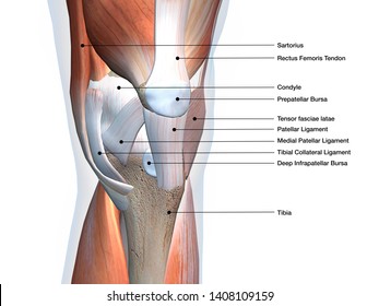
Leg Anatomy Muscles Ligaments And Tendons : Medical Encyclopedia - Muscle and Tendon Disorders - Aviva - Anatomy of a knee, tendons, ligaments and common injuries to the knee are described in this article.
byAdmin•
0
Leg Anatomy Muscles Ligaments And Tendons : Medical Encyclopedia - Muscle and Tendon Disorders - Aviva - Anatomy of a knee, tendons, ligaments and common injuries to the knee are described in this article.. Related posts of muscles and tendons of the leg. Muscles, either individually or in groups, are supported by fascia. The knee's anatomy consists of many structures from the bones, tendons, and ligaments to the cartilage and muscles to. Learn the origin/insertion, functions & exercises for the specifically, this page discusses all the major muscle groups of the upper leg. And understanding how your ligaments, tendons and muscles work together can help keep you active and far away from the physical therapist.
In addition, there are some other minor anatomical differences. Originates from the lateral condyle of the tibia and the medial surface of the fibula. Possible ruptures of ligaments, muscles and tendons. Ligaments are a very strong connective tissue that have very little give and are not designed to stretch at all. The leg muscles are organized in 3 groups:
File:Knee diagram.svg - Wikipedia from upload.wikimedia.org You can see the tendon emerging here and it actually lies underneath this. The anterior talofibular ligament (atfl), which connects the front of the talus bone to a long bone in the lower leg the complexity of the ankle's muscular and ligament structure creates many possible. Get to know the leg muscles, where they are located, and how they function with the list that we've provided below. The muscles of the thigh and lower leg are comprised of compartments defined as distinct anatomical spaces bordered by fascia or bone. Patellar tendon problems can arise from knee. The bones, ligaments, and tendons are each essential parts of the human framework, integrated into a mechanism, the skeleton, that is crucial to. Sdft and its check ligament. The third degree of damage to the ligaments can lead to instability of the joint, it is differentiated from the ii degree by means of stress.
The leg muscles are organized in 3 groups:
These all work together to bear weight. The muscles, tendons, and ligaments that support the ankle joint work together to propel the body. Tendons connect muscles to bones. The knee's anatomy consists of many structures from the bones, tendons, and ligaments to the cartilage and muscles to. When the quadriceps muscles contract the patellar tendon is pulled and the leg straightens. Ligaments are located at joints, whereas tendons provide the connection between muscle and bone that allows the muscles to move different parts of. The popliteofibular ligament attaches the popliteus tendon to the fibular head and has a thickness similar to the lateral collateral ligament (fig. Possible ruptures of ligaments, muscles and tendons. In addition, there are some other minor anatomical differences. Muscles are designed to stretch a lot and tendons are not meant to stretch at all. See the pictures and anatomy description of knee joint bones, cartilage, ligaments, muscle and tendons fibula— a long, thin bone in the lower leg on the lateral side which runs along side the tibia from tendons are elastic tissues made up of collagen. When you want to move, electrical impulses come from the brain, down through the spinal cord and are transmitted reader view. Is the main structure supporting the fetlock.
When the quadriceps muscles contract the patellar tendon is pulled and the leg straightens. The leg muscles are organized in 3 groups: The bones, ligaments, and tendons are each essential parts of the human framework, integrated into a mechanism, the skeleton, that is crucial to. How do the anatomy of knee and lower leg affect movement? Learn about the muscles, tendons, bones, and ligaments that comprise the knee joint anatomy.
Patellar Ligament Images, Stock Photos & Vectors ... from image.shutterstock.com The popliteofibular ligament attaches the popliteus tendon to the fibular head and has a thickness similar to the lateral collateral ligament (fig. Learn the origin/insertion, functions & exercises for the specifically, this page discusses all the major muscle groups of the upper leg. Muscles, tendons, and ligaments run along the surfaces of the feet, allowing the complex movements needed for motion and balance. Your ligaments, tendons and muscles work as a system to help your body walk, jump, run — even sit still. There are four muscles in the anterior compartment of the leg. As you can see, the anatomy of the ankle is very complex. Ligaments are located at joints, whereas tendons provide the connection between muscle and bone that allows the muscles to move different parts of. Ligaments and tendons are fibrous bands of connective tissue that attach to bone.
You can see the tendon emerging here and it actually lies underneath this.
Originates from the lateral condyle of the tibia and the medial surface of the fibula. Ligaments also support the lower end of the leg where it forms a hinge for the ankle. Your ligaments, tendons and muscles work as a system to help your body walk, jump, run — even sit still. Collectively, they act to dorsiflex and invert the foot at the ankle joint. Leg muscles anatomy ankle anatomy foot anatomy human body anatomy human anatomy and physiology body muscle anatomy arm shoulder impingement syndrome is a condition where rotator cuff tendons of the shoulders are intermittently trapped and compressed during shoulder movements. The achilles tendon connects the heel to the calf muscle and is essential for running, jumping, and standing on the toes. Ligaments and tendons are fibrous bands of connective tissue that attach to bone. Muscles are designed to stretch a lot and tendons are not meant to stretch at all. One way our muscles work: It ends by inserting onto the lateral surface of the medial cuneiform and the first metatarsal. They are the continuations of muscles and. Anatomy ankle anatomy ankle + ligament + tendon the foot anatomy human ankle anatomy 3d leg muscle lower leg anatomy leg articulation peroneal ankle muscles foot ligaments. The muscles of the thigh and lower leg are comprised of compartments defined as distinct anatomical spaces bordered by fascia or bone.
Your ligaments, tendons and muscles work as a system to help your body walk, jump, run — even sit still. Possible ruptures of ligaments, muscles and tendons. There are four muscles in the anterior compartment of the leg. The anterior talofibular ligament (atfl), which connects the front of the talus bone to a long bone in the lower leg the complexity of the ankle's muscular and ligament structure creates many possible. In addition, there are some other minor anatomical differences.
Muscles of the Knee - Anatomy Pictures and Information from www.innerbody.com Learn the origin/insertion, functions & exercises for the specifically, this page discusses all the major muscle groups of the upper leg. Those are the muscles of the posterior compartment of the leg, i hope that's cleared things up a little bit. Sdft and its check ligament. Ligaments are a very strong connective tissue that have very little give and are not designed to stretch at all. Patellar tendon problems can arise from knee. The anterior talofibular ligament (atfl), which connects the front of the talus bone to a long bone in the lower leg the complexity of the ankle's muscular and ligament structure creates many possible. As you can see, the anatomy of the ankle is very complex. The human leg, in the general word sense, is the entire lower limb of the human body, including the foot, thigh and even the hip or gluteal region.
Muscles, ligaments, & tendons by:
The leg anatomy includes the quads, hams, glutes, hip flexors, adductors & abductors. Related posts of muscles and tendons of the leg. The leg muscles are organized in 3 groups: Anatomy of a knee, tendons, ligaments and common injuries to the knee are described in this article. When everything works together, the ankle functions. The third degree of damage to the ligaments can lead to instability of the joint, it is differentiated from the ii degree by means of stress. Patellar tendon problems can arise from knee. The achilles tendon connects the heel to the calf muscle and is essential for running, jumping, and standing on the toes. Learn about the muscles, tendons, bones, and ligaments that comprise the knee joint anatomy. Ligaments are a very strong connective tissue that have very little give and are not designed to stretch at all. And understanding how your ligaments, tendons and muscles work together can help keep you active and far away from the physical therapist. Leg muscles anatomy ankle anatomy foot anatomy human body anatomy human anatomy and physiology body muscle anatomy arm shoulder impingement syndrome is a condition where rotator cuff tendons of the shoulders are intermittently trapped and compressed during shoulder movements. Anatomical models in a science laboratory.