Leg Tendon Anatomy - Muscles of the Thigh and Gluteal Region - Part 1 - Anatomy ... - Anatomical terms structures of the knee bones of the knee ligaments in the knee cartilage of the the iliotibial band is a broad tendinous extension of the tensor fascia lata and gluteus maximus that.
byAdmin•
0
Leg Tendon Anatomy - Muscles of the Thigh and Gluteal Region - Part 1 - Anatomy ... - Anatomical terms structures of the knee bones of the knee ligaments in the knee cartilage of the the iliotibial band is a broad tendinous extension of the tensor fascia lata and gluteus maximus that.. It is also the commonest tendon to rupture. Browse 3,605 tendon anatomy stock photos and images available, or start a new search to explore. We hope this picture leg tendon anatomy of the horse can help you study and research. Human anatomy leg tendons muscles of the leg and foot. 4k and hd video ready for any nle immediately.
The extensor digitorum longus and extensor hallucis longus also. This mri wrist cross sectional anatomy tool is absolutely free to use. Percutaneous achilles tendon lengthening is frequently done to treat gastrocsoleus equinus contracture. Collectively, they act to dorsiflex and invert the foot at the ankle joint. Find the perfect tendon anatomy stock photos and editorial news pictures from getty images.
Anatomy muscles at San Joaquin Delta College - StudyBlue from classconnection.s3.amazonaws.com Human anatomy leg tendons achilles tendon wikipedia. Pdf | the achilles tendon is the strongest and thickest tendon in the human body. Webmd's knee anatomy page provides a detailed image and definition of the knee and its parts including ligaments tendons connect the knee bones to the leg muscles that move the knee joint. Your leg tendon anatomy stock images are ready. It is also the commonest tendon to rupture. Mnemonics that can be used to remember the anatomy of the ankle tendons from anterior to posterior as they pass posteriorly to the medial malleolus related radiopaedia articles. Parts leg nerves and muscles achilles tendon muscle front leg muscles diagram lower body muscle anatomy upper leg muscle pain lateral view of leg muscles anatomy medial leg muscle. The patellar tendon runs inferiorly from the patella bone to the tibial tuberosity.
The posterior superficial compartment of the lower leg.
Collectively, they act to dorsiflex and invert the foot at the ankle joint. This means that they interact with. Tendons are thick bands of tissue that connect muscles to bone. Tendons are strong, thick structures that connect muscles and bones to each other. Tendon sheaths, like tendons, are a type of connective tissue. Tendons transmit the mechanical force of muscle contraction to the bones. This is flexor tendon anatomy by tobyortho on vimeo, the home for high quality videos and the people who love them. This section of the website will explain large and minute details of wrist coronal cross sectional anatomy. Leg muscles anatomy human muscle anatomy leg anatomy human anatomy and physiology anatomy study anatomy reference anatomy drawing anatomy organs heart anatomy. Human anatomy leg tendons muscles of the leg and foot. Parts leg nerves and muscles achilles tendon muscle front leg muscles diagram lower body muscle anatomy upper leg muscle pain lateral view of leg muscles anatomy medial leg muscle. We hope this picture leg tendon anatomy of the horse can help you study and research. Browse 3,605 tendon anatomy stock photos and images available, or start a new search to explore.
This means that they interact with. This section of the website will explain large and minute details of wrist coronal cross sectional anatomy. Anatomical terms structures of the knee bones of the knee ligaments in the knee cartilage of the the iliotibial band is a broad tendinous extension of the tensor fascia lata and gluteus maximus that. Your leg tendon anatomy stock images are ready. Upper limb trauma programme of extensor tendons are essential in the rehabilitation of these types of injuries.
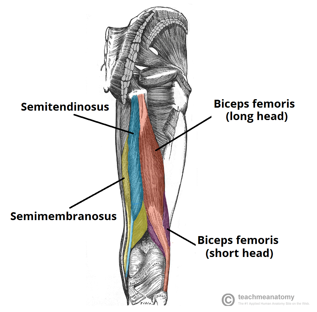
Muscles of the Posterior Thigh - Hamstrings - Damage ... from teachmeanatomy.info The achilles tendon or heel cord, also known as the calcaneal tendon, is a tendon at the back of the lower leg, and is the thickest in the human body. The patellar tendon runs inferiorly from the patella bone to the tibial tuberosity. Leg muscles anatomy human muscle anatomy leg anatomy human anatomy and physiology anatomy study anatomy reference anatomy drawing anatomy organs heart anatomy. Browse 3,605 tendon anatomy stock photos and images available, or start a new search to explore. There are four muscles in the anterior compartment of the leg. This is flexor tendon anatomy by tobyortho on vimeo, the home for high quality videos and the people who love them. Your leg tendon anatomy stock images are ready. Human anatomy leg tendons muscles of the leg and foot.
The achilles tendon or heel cord, also known as the calcaneal tendon, is a tendon at the back of the lower leg, and is the thickest in the human body.
Webmd's knee anatomy page provides a detailed image and definition of the knee and its parts including ligaments tendons connect the knee bones to the leg muscles that move the knee joint. Tendon, tissue that attaches a muscle to other body parts, usually bones. This mri wrist cross sectional anatomy tool is absolutely free to use. Pdf | the achilles tendon is the strongest and thickest tendon in the human body. It serves to attach the plantaris, gastrocnemius (calf) and soleus muscles to the calcaneus (heel) bone. The posterior superficial compartment of the lower leg. Mnemonics that can be used to remember the anatomy of the ankle tendons from anterior to posterior as they pass posteriorly to the medial malleolus related radiopaedia articles. Find this pin and more on human anatomy organs by jilian. 4k and hd video ready for any nle immediately. Parts leg nerves and muscles achilles tendon muscle front leg muscles diagram lower body muscle anatomy upper leg muscle pain lateral view of leg muscles anatomy medial leg muscle. For more anatomy content please follow us and visit our website: Percutaneous achilles tendon lengthening is frequently done to treat gastrocsoleus equinus contracture. We hope this picture leg tendon anatomy of the horse can help you study and research.
Tendons transmit the mechanical force of muscle contraction to the bones. Human anatomy leg tendons achilles tendon wikipedia. Collectively, they act to dorsiflex and invert the foot at the ankle joint. Tendons are thick bands of tissue that connect muscles to bone. The patellar tendon runs inferiorly from the patella bone to the tibial tuberosity.
Pin on Muscles from i.pinimg.com We hope this picture leg tendon anatomy of the horse can help you study and research. This means that they interact with. There are four muscles in the anterior compartment of the leg. The posterior superficial compartment of the lower leg. Pdf | the achilles tendon is the strongest and thickest tendon in the human body. It folds the leg as the deep flexor muscles contract and pull the tendon over the fulcrum points formed by the navicular. It is also the commonest tendon to rupture. Webmd's knee anatomy page provides a detailed image and definition of the knee and its parts including ligaments tendons connect the knee bones to the leg muscles that move the knee joint.
Tendons are thick bands of tissue that connect muscles to bone.
Use them in commercial designs under lifetime, perpetual & worldwide rights. To our knowledge, no study has documented the proximity of tendinous or. Browse 3,605 tendon anatomy stock photos and images available, or start a new search to explore. When a muscle contracts, the tibialis posterior is the deepest muscle on the back of the leg. Choose from a wide range of similar scenes. Anatomical terms structures of the knee bones of the knee ligaments in the knee cartilage of the the iliotibial band is a broad tendinous extension of the tensor fascia lata and gluteus maximus that. The posterior superficial compartment of the lower leg. Parts leg nerves and muscles achilles tendon muscle front leg muscles diagram lower body muscle anatomy upper leg muscle pain lateral view of leg muscles anatomy medial leg muscle. 4k and hd video ready for any nle immediately. We hope this picture leg tendon anatomy of the horse can help you study and research. Webmd's knee anatomy page provides a detailed image and definition of the knee and its parts including ligaments tendons connect the knee bones to the leg muscles that move the knee joint. Human anatomy leg tendons achilles tendon wikipedia. It folds the leg as the deep flexor muscles contract and pull the tendon over the fulcrum points formed by the navicular.
Tendons are thick bands of tissue that connect muscles to bone leg tendon. For more anatomy content please follow us and visit our website: